Review


polyclonal anti versican antibody  (OriGene)


Bioz Verified Symbol OriGene is a verified supplier
Bioz Manufacturer Symbol OriGene manufactures this product  
  • Logo
  • About
  • News
  • Press Release
  • Team
  • Advisors
  • Partners
  • Contact
  • Bioz Stars
  • Bioz vStars
  • 90

    Structured Review

    OriGene polyclonal anti versican antibody
    Polyclonal Anti Versican Antibody, supplied by OriGene, used in various techniques. Bioz Stars score: 90/100, based on 2 PubMed citations. ZERO BIAS - scores, article reviews, protocol conditions and more
    https://www.bioz.com/product/polyclonal+rabbit+anti+versican/Versican+(VCAN)+Rabbit+Polyclonal+Antibody/us12011499-312-6-9
    Average 90 stars, based on 2 article reviews
    polyclonal anti versican antibody - by Bioz Stars, 2026-09
    90/100 stars

    Images

    Related Articles

    Cell Culture:

    Article Title: NG2 and phosphacan are present in the astroglial scar after human traumatic spinal cord injury
    Article Snippet: .. The primary antibodies used were: monoclonal mouse anti-human NG2 (clone B5, undiluted cell culture supernatant from ATCC cultures and antibody 9.2.27, diluted 1:50; gift from Prof. R. Reisfeld, Scripps Research Institute), monoclonal mouse anti-neurocan (Chemicon, diluted 1:100), polyclonal rabbit anti-versican (Acris antibodies, diluted 1:1.000), monoclonal mouse anti-phosphacan (Chemicon, diluted 1:500), polyclonal rabbit anti-GFAP (DAKO, diluted 1:2.500), polyclonal rabbit anti-myelin basic protein (MBP) (Chemicon, diluted 1:1.000) and polyclonal rabbit anti-neurofilament (NF, Sigma-Aldrich, diluted 1:2.000). .. Following extensive rinsing steps in 0.1 M PBS, sections were incubated in biotinylated horse anti-mouse or anti-rabbit antibody (Vector Laboratories, diluted 1:500) for 1 hour at room temperature.


    Similar Products

    90
    OriGene polyclonal anti versican antibody
    Polyclonal Anti Versican Antibody, supplied by OriGene, used in various techniques. Bioz Stars score: 90/100, based on 1 PubMed citations. ZERO BIAS - scores, article reviews, protocol conditions and more
    https://www.bioz.com/product/polyclonal+rabbit+anti+versican/Versican+(VCAN)+Rabbit+Polyclonal+Antibody/us12011499-312-6-9
    Average 90 stars, based on 1 article reviews
    polyclonal anti versican antibody - by Bioz Stars, 2026-09
    90/100 stars
      Buy from Supplier

    92
    Bioss bs 2533r rabbit igg polyclonal tnc
    Bs 2533r Rabbit Igg Polyclonal Tnc, supplied by Bioss, used in various techniques. Bioz Stars score: 92/100, based on 1 PubMed citations. ZERO BIAS - scores, article reviews, protocol conditions and more
    https://www.bioz.com/product/polyclonal+rabbit+anti+versican/Versican+Polyclonal+Antibody/pmc10520636__ADVS___10___2301930___s001-73-35-34
    Average 92 stars, based on 1 article reviews
    bs 2533r rabbit igg polyclonal tnc - by Bioz Stars, 2026-09
    92/100 stars
      Buy from Supplier

    92
    Bioss bs 2533r rabbit igg polyclonal vcan human
    Bs 2533r Rabbit Igg Polyclonal Vcan Human, supplied by Bioss, used in various techniques. Bioz Stars score: 92/100, based on 1 PubMed citations. ZERO BIAS - scores, article reviews, protocol conditions and more
    https://www.bioz.com/product/polyclonal+rabbit+anti+versican/Versican+Polyclonal+Antibody/pmc10520636__ADVS___10___2301930___s001-71-116-115
    Average 92 stars, based on 1 article reviews
    bs 2533r rabbit igg polyclonal vcan human - by Bioz Stars, 2026-09
    92/100 stars
      Buy from Supplier

    86
    Danaher Inc rabbit polyclonal anti versican
    Rabbit Polyclonal Anti Versican, supplied by Danaher Inc, used in various techniques. Bioz Stars score: 86/100, based on 1 PubMed citations. ZERO BIAS - scores, article reviews, protocol conditions and more
    https://www.bioz.com/product/polyclonal+rabbit+anti+versican/pm37174615-90-54-49
    Average 86 stars, based on 1 article reviews
    rabbit polyclonal anti versican - by Bioz Stars, 2026-09
    86/100 stars
      Buy from Supplier

    92
    Novus Biologicals anti vcan rabbit polyclonal antibody
    Inhibition of miR-124-3p expression rescues at RA-induced CP in mice. (A) Schematic experimental design. (B) Gross picture of E18.5 C57BL/6J mice treated with control vehicle, at RA, or at RA + miR-124-3p inhibitor. Bars, 1 mm. (C) H&E staining of the face of E18.5 C57BL/6J mice treated with control vehicle, at RA, or at RA + miR-124-3p inhibitor. Bars, 400 μm. (D) Ki-67 staining (green) in the developing palate of E13.5 C57BL/6J mice treated with/without a miR-124-3p inhibitor under at RA condition. Nuclei were counterstained with DAPI (blue). Representative images from two independent experiments are shown. Graph shows the quantification of Ki-67 positive cells. One-way ANOVA with Tukey’s honest significant difference test. ** p < 0.01 vs. control. ## p < 0.01 vs. at RA. Bars, 50 μm. (E) Quantitative RT-PCR for the indicated genes after treatment with/without a miR-124-3p inhibitor under at RA condition in the palatal shelves of E14.5 C57BL/6J mice. Two-way ANOVA with Tukey’s honest significant difference test ( n = 3). *Adjusted p < 0.00313 (16 genes), **adjusted p < 0.000625 (16 genes), and ***adjusted p < 0.0000625 (16 genes) vs. control. # Adjusted p < 0.00313 (16 genes), ## adjusted p < 0.000625 (16 genes), and ### adjusted p < 0.0000625 (16 genes) vs. at RA. (F) AZAN staining (upper panels) and immunohistochemical staining for <t>VCAN</t> (middle panels; brown) and CDC42 (bottom panels; brown) in E13.5 C57BL/6J mice treated with control vehicle, at RA, or at RA + miR-124-3p inhibitor. Bars, 25 μm.
    Anti Vcan Rabbit Polyclonal Antibody, supplied by Novus Biologicals, used in various techniques. Bioz Stars score: 92/100, based on 1 PubMed citations. ZERO BIAS - scores, article reviews, protocol conditions and more
    https://www.bioz.com/product/polyclonal+rabbit+anti+versican/Versican+Antibody/pmc08219963-74-42-46
    Average 92 stars, based on 1 article reviews
    anti vcan rabbit polyclonal antibody - by Bioz Stars, 2026-09
    92/100 stars
      Buy from Supplier

    90
    Millipore polyclonal rabbit anti versican

    Polyclonal Rabbit Anti Versican, supplied by Millipore, used in various techniques. Bioz Stars score: 90/100, based on 1 PubMed citations. ZERO BIAS - scores, article reviews, protocol conditions and more
    https://www.bioz.com/product/polyclonal+rabbit+anti+versican/polyclonal+rabbit+anti+versican+antibody++%CE%B2+gag/pmc08215304-4-0-5
    Average 90 stars, based on 1 article reviews
    polyclonal rabbit anti versican - by Bioz Stars, 2026-09
    90/100 stars
      Buy from Supplier

    90
    Millipore rabbit anti-mouse versican (gag beta domain) polyclonal antibody
    Representative Raman images and immunofluorescence (IF) staining and histochemistry for WT, fibulin-5 knockout ( Fbln5 KO ), and smooth muscle-specific fibulin-4 knockout ( Fbln4 SMKO ) mice (A) Raman images of large-area scans. Cross-sections of the ascending aorta from WT, Fbln5 KO , and Fbln4 SMKO . False-color intensity distribution heatmaps for elastic fibers (green), collagen fibers (red), nuclei (blue), aggrecan (yellow), <t>versican</t> (pink), lipids (orange), and residual ECM (cyan). Scale bars, 50 μm. see also <xref ref-type=Figure S1 . (B) Gross photos of murine aortas of WT, Fbln5 KO , and Fbln4 SMKO mice. Fbln5 KO exhibits elongation of the ascending aorta and tortuous descending aorta. Fbln4 SMKO shows a large aneurysm in the ascending aorta but not in the descending aorta. Scale bars, 1 mm. (C) IF staining for collagen type I, aggrecan, and versican in red, elastin autofluorescence (green), and nuclei (blue). Scale bars, 20 μm. (D) Routine histochemical staining for Alcian blue (glycosaminoglycans) and oil red O (lipid) staining. Scale bars, 20 μm. L, luminal side. " width="250" height="auto" />
    Rabbit Anti Mouse Versican (Gag Beta Domain) Polyclonal Antibody, supplied by Millipore, used in various techniques. Bioz Stars score: 90/100, based on 1 PubMed citations. ZERO BIAS - scores, article reviews, protocol conditions and more
    https://www.bioz.com/product/polyclonal+rabbit+anti+versican/rabbit+anti+mouse+versican+%CE%B2+gag+domain/pmc08149374-3-0-9
    Average 90 stars, based on 1 article reviews
    rabbit anti-mouse versican (gag beta domain) polyclonal antibody - by Bioz Stars, 2026-09
    90/100 stars
      Buy from Supplier

    90
    Millipore rabbit polyclonal anti-mouse versican gag β domain
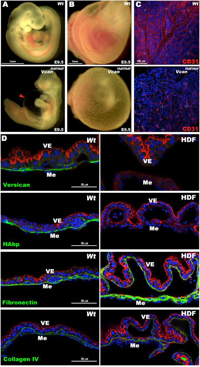
    (A) E9.5 wild type and Vcan hdf/hdf embryos. The red arrowhead shows the dilated pericardial sac in the latter. (B) E9.5 yolk sacs imaged in situ demonstrates the absence of vasculature in the E9.5 Vcan hdf/hdf yolk sac. (C) Three-dimensional (3D) maximum-intensity projections of whole-mount yolk sacs stained with anti-CD31 (red) showing absence of the vascular network in E9.5 Vcan hdf/hdf yolk sacs (n=3 yolk sacs of each genotype). (D) <t>Versican</t> <t>GAG</t> β staining (green) is present throughout the mesoderm (Me) of E9.5 wild type yolk sacs and absent in the Vcan hdf/hdf yolk sac. F-actin (red) staining highlights visceral endoderm (VE), detaches from mesoderm in Vcan hdf/hdf yolk sacs. HA and fibronectin (green) were similarly distributed as versican in wild type yolk sac, but HA staining was absent in Vcan hdf/hdf yolk sac and fibronectin staining was more intense. Collagen IV staining intensity was similar in wild type and Vcan hdf/hdf yolk sacs (n=3 yolk sacs of each genotype). Scale bar in A, B=1mm, C= 100μm, D= 50μm.
    Rabbit Polyclonal Anti Mouse Versican Gag β Domain, supplied by Millipore, used in various techniques. Bioz Stars score: 90/100, based on 1 PubMed citations. ZERO BIAS - scores, article reviews, protocol conditions and more
    https://www.bioz.com/product/polyclonal+rabbit+anti+versican/versican+antibody/bio_rxiv__753418-211-23-30
    Average 90 stars, based on 1 article reviews
    rabbit polyclonal anti-mouse versican gag β domain - by Bioz Stars, 2026-09
    90/100 stars
      Buy from Supplier

    90
    Millipore antibody rabbit polyclonal anti-versican gag-beta

    Antibody Rabbit Polyclonal Anti Versican Gag Beta, supplied by Millipore, used in various techniques. Bioz Stars score: 90/100, based on 1 PubMed citations. ZERO BIAS - scores, article reviews, protocol conditions and more
    https://www.bioz.com/product/polyclonal+rabbit+anti+versican/rabbit+polyclonal+anti+versican+gag%CE%B2+domain+antibody/pmc07529456-23-2-7
    Average 90 stars, based on 1 article reviews
    antibody rabbit polyclonal anti-versican gag-beta - by Bioz Stars, 2026-09
    90/100 stars
      Buy from Supplier

    Image Search Results


    Inhibition of miR-124-3p expression rescues at RA-induced CP in mice. (A) Schematic experimental design. (B) Gross picture of E18.5 C57BL/6J mice treated with control vehicle, at RA, or at RA + miR-124-3p inhibitor. Bars, 1 mm. (C) H&E staining of the face of E18.5 C57BL/6J mice treated with control vehicle, at RA, or at RA + miR-124-3p inhibitor. Bars, 400 μm. (D) Ki-67 staining (green) in the developing palate of E13.5 C57BL/6J mice treated with/without a miR-124-3p inhibitor under at RA condition. Nuclei were counterstained with DAPI (blue). Representative images from two independent experiments are shown. Graph shows the quantification of Ki-67 positive cells. One-way ANOVA with Tukey’s honest significant difference test. ** p < 0.01 vs. control. ## p < 0.01 vs. at RA. Bars, 50 μm. (E) Quantitative RT-PCR for the indicated genes after treatment with/without a miR-124-3p inhibitor under at RA condition in the palatal shelves of E14.5 C57BL/6J mice. Two-way ANOVA with Tukey’s honest significant difference test ( n = 3). *Adjusted p < 0.00313 (16 genes), **adjusted p < 0.000625 (16 genes), and ***adjusted p < 0.0000625 (16 genes) vs. control. # Adjusted p < 0.00313 (16 genes), ## adjusted p < 0.000625 (16 genes), and ### adjusted p < 0.0000625 (16 genes) vs. at RA. (F) AZAN staining (upper panels) and immunohistochemical staining for VCAN (middle panels; brown) and CDC42 (bottom panels; brown) in E13.5 C57BL/6J mice treated with control vehicle, at RA, or at RA + miR-124-3p inhibitor. Bars, 25 μm.

    Journal: Frontiers in Cell and Developmental Biology

    Article Title: MicroRNA-124-3p Plays a Crucial Role in Cleft Palate Induced by Retinoic Acid

    doi: 10.3389/fcell.2021.621045

    Figure Lengend Snippet: Inhibition of miR-124-3p expression rescues at RA-induced CP in mice. (A) Schematic experimental design. (B) Gross picture of E18.5 C57BL/6J mice treated with control vehicle, at RA, or at RA + miR-124-3p inhibitor. Bars, 1 mm. (C) H&E staining of the face of E18.5 C57BL/6J mice treated with control vehicle, at RA, or at RA + miR-124-3p inhibitor. Bars, 400 μm. (D) Ki-67 staining (green) in the developing palate of E13.5 C57BL/6J mice treated with/without a miR-124-3p inhibitor under at RA condition. Nuclei were counterstained with DAPI (blue). Representative images from two independent experiments are shown. Graph shows the quantification of Ki-67 positive cells. One-way ANOVA with Tukey’s honest significant difference test. ** p < 0.01 vs. control. ## p < 0.01 vs. at RA. Bars, 50 μm. (E) Quantitative RT-PCR for the indicated genes after treatment with/without a miR-124-3p inhibitor under at RA condition in the palatal shelves of E14.5 C57BL/6J mice. Two-way ANOVA with Tukey’s honest significant difference test ( n = 3). *Adjusted p < 0.00313 (16 genes), **adjusted p < 0.000625 (16 genes), and ***adjusted p < 0.0000625 (16 genes) vs. control. # Adjusted p < 0.00313 (16 genes), ## adjusted p < 0.000625 (16 genes), and ### adjusted p < 0.0000625 (16 genes) vs. at RA. (F) AZAN staining (upper panels) and immunohistochemical staining for VCAN (middle panels; brown) and CDC42 (bottom panels; brown) in E13.5 C57BL/6J mice treated with control vehicle, at RA, or at RA + miR-124-3p inhibitor. Bars, 25 μm.

    Article Snippet: After antigen retrieval treatment with citrate buffer (pH 6.0) and blocking of endogenous peroxidase with 0.3% hydrogen peroxide in methanol, the sections were incubated with anti-cytokeratin 14 mouse monoclonal antibody (Abcam, ab7800, 1:200 dilution), anti-Ki-67 rabbit monoclonal antibody (Abcam, ab16667, 1:200 dilution), anti-VCAN rabbit polyclonal antibody (Novus Biologicals, Centennial, CO, United States, NBP1-85432, 1:200 dilution), or anti-CDC42 rabbit polyclonal antibody (Proteintech, Rosemont, IL, United States, 10155-1-AP, 1:50 dilution) at 4°C overnight.

    Techniques: Inhibition, Expressing, Control, Staining, Quantitative RT-PCR, Immunohistochemical staining

    Journal: iScience

    Article Title: Modelling TGFβR and Hh pathway regulation of prognostic matrisome molecules in ovarian cancer

    doi: 10.1016/j.isci.2021.102674

    Figure Lengend Snippet:

    Article Snippet: Polyclonal Rabbit Anti Versican , Sigma , Cat# HPA004726; RRID: AB_1080561.

    Techniques: Activation Assay, Plasmid Preparation, Formalin-fixed Paraffin-Embedded, Recombinant, Enzyme-linked Immunosorbent Assay, Isolation, Software

    Representative Raman images and immunofluorescence (IF) staining and histochemistry for WT, fibulin-5 knockout ( Fbln5 KO ), and smooth muscle-specific fibulin-4 knockout ( Fbln4 SMKO ) mice (A) Raman images of large-area scans. Cross-sections of the ascending aorta from WT, Fbln5 KO , and Fbln4 SMKO . False-color intensity distribution heatmaps for elastic fibers (green), collagen fibers (red), nuclei (blue), aggrecan (yellow), versican (pink), lipids (orange), and residual ECM (cyan). Scale bars, 50 μm. see also <xref ref-type=Figure S1 . (B) Gross photos of murine aortas of WT, Fbln5 KO , and Fbln4 SMKO mice. Fbln5 KO exhibits elongation of the ascending aorta and tortuous descending aorta. Fbln4 SMKO shows a large aneurysm in the ascending aorta but not in the descending aorta. Scale bars, 1 mm. (C) IF staining for collagen type I, aggrecan, and versican in red, elastin autofluorescence (green), and nuclei (blue). Scale bars, 20 μm. (D) Routine histochemical staining for Alcian blue (glycosaminoglycans) and oil red O (lipid) staining. Scale bars, 20 μm. L, luminal side. " width="100%" height="100%">

    Journal: Cell Reports Medicine

    Article Title: Raman microspectroscopy and Raman imaging reveal biomarkers specific for thoracic aortic aneurysms

    doi: 10.1016/j.xcrm.2021.100261

    Figure Lengend Snippet: Representative Raman images and immunofluorescence (IF) staining and histochemistry for WT, fibulin-5 knockout ( Fbln5 KO ), and smooth muscle-specific fibulin-4 knockout ( Fbln4 SMKO ) mice (A) Raman images of large-area scans. Cross-sections of the ascending aorta from WT, Fbln5 KO , and Fbln4 SMKO . False-color intensity distribution heatmaps for elastic fibers (green), collagen fibers (red), nuclei (blue), aggrecan (yellow), versican (pink), lipids (orange), and residual ECM (cyan). Scale bars, 50 μm. see also Figure S1 . (B) Gross photos of murine aortas of WT, Fbln5 KO , and Fbln4 SMKO mice. Fbln5 KO exhibits elongation of the ascending aorta and tortuous descending aorta. Fbln4 SMKO shows a large aneurysm in the ascending aorta but not in the descending aorta. Scale bars, 1 mm. (C) IF staining for collagen type I, aggrecan, and versican in red, elastin autofluorescence (green), and nuclei (blue). Scale bars, 20 μm. (D) Routine histochemical staining for Alcian blue (glycosaminoglycans) and oil red O (lipid) staining. Scale bars, 20 μm. L, luminal side.

    Article Snippet: Rabbit anti-mouse versican (GAG beta domain) polyclonal antibody , Millipore , Cat#: AB1033, RRID: AB_90462.

    Techniques: Immunofluorescence, Staining, Knock-Out

    Journal: Cell Reports Medicine

    Article Title: Raman microspectroscopy and Raman imaging reveal biomarkers specific for thoracic aortic aneurysms

    doi: 10.1016/j.xcrm.2021.100261

    Figure Lengend Snippet:

    Article Snippet: Rabbit anti-mouse versican (GAG beta domain) polyclonal antibody , Millipore , Cat#: AB1033, RRID: AB_90462.

    Techniques: Plasmid Preparation, Blocking Assay, Recombinant, Software

    (A) E9.5 wild type and Vcan hdf/hdf embryos. The red arrowhead shows the dilated pericardial sac in the latter. (B) E9.5 yolk sacs imaged in situ demonstrates the absence of vasculature in the E9.5 Vcan hdf/hdf yolk sac. (C) Three-dimensional (3D) maximum-intensity projections of whole-mount yolk sacs stained with anti-CD31 (red) showing absence of the vascular network in E9.5 Vcan hdf/hdf yolk sacs (n=3 yolk sacs of each genotype). (D) Versican GAG β staining (green) is present throughout the mesoderm (Me) of E9.5 wild type yolk sacs and absent in the Vcan hdf/hdf yolk sac. F-actin (red) staining highlights visceral endoderm (VE), detaches from mesoderm in Vcan hdf/hdf yolk sacs. HA and fibronectin (green) were similarly distributed as versican in wild type yolk sac, but HA staining was absent in Vcan hdf/hdf yolk sac and fibronectin staining was more intense. Collagen IV staining intensity was similar in wild type and Vcan hdf/hdf yolk sacs (n=3 yolk sacs of each genotype). Scale bar in A, B=1mm, C= 100μm, D= 50μm.

    Journal: bioRxiv

    Article Title: The versican-hyaluronan complex provides an essential extracellular matrix niche for Flk1+ hematoendothelial progenitors

    doi: 10.1101/753418

    Figure Lengend Snippet: (A) E9.5 wild type and Vcan hdf/hdf embryos. The red arrowhead shows the dilated pericardial sac in the latter. (B) E9.5 yolk sacs imaged in situ demonstrates the absence of vasculature in the E9.5 Vcan hdf/hdf yolk sac. (C) Three-dimensional (3D) maximum-intensity projections of whole-mount yolk sacs stained with anti-CD31 (red) showing absence of the vascular network in E9.5 Vcan hdf/hdf yolk sacs (n=3 yolk sacs of each genotype). (D) Versican GAG β staining (green) is present throughout the mesoderm (Me) of E9.5 wild type yolk sacs and absent in the Vcan hdf/hdf yolk sac. F-actin (red) staining highlights visceral endoderm (VE), detaches from mesoderm in Vcan hdf/hdf yolk sacs. HA and fibronectin (green) were similarly distributed as versican in wild type yolk sac, but HA staining was absent in Vcan hdf/hdf yolk sac and fibronectin staining was more intense. Collagen IV staining intensity was similar in wild type and Vcan hdf/hdf yolk sacs (n=3 yolk sacs of each genotype). Scale bar in A, B=1mm, C= 100μm, D= 50μm.

    Article Snippet: For staining whole mount embryos, yolk sacs, vibratome sections and cell culture chamber slides the following antibodies, staining reagents and dilutions were used: Rabbit polyclonal anti-mouse versican GAG β domain (Millipore-Sigma, catalog no. AB1033) 1:200; Rabbit polyclonal anti-versican cleavage site (anti-Vc) [ ] 1:400; Rabbit polyclonal anti-versican V 0 /V 1 neo cleavage antibody (DPEAAE) (Thermo Fisher, catalog no. PA1-1748A) 1:400; biotinylated hyaluronan binding protein (HAbp) (Millipore-Sigma, Calbiochem, catalog no. 385911) 1:100; rabbit polyclonal anti-fibronectin (Abcam, catalog no. Ab2413) 1:200; rabbit polyclonal anti-collagen-IV (Rockland antibodies and assays, catalog no. 600-401-106) 1:400; rat monoclonal anti-mouse Flk1 (clone Avas12, Thermo Fisher, catalog no. 17-5821-81) 1:200; hamster anti-mouse CD31 (Milipore-Sigma, catalog no. MAB1398Z) 1:400; FITC-conjugated anti-mouse CD41 (Biolegend, catalog no.133904) 1:100; mouse monoclonal Cy3 conjugated smooth muscle α -actin (Sigma-Aldrich, catalog no. C6198) 1:600;Alexa Fluor-568 phalloidin (Life Technologies, catalog no. A12380) 1:500; Streptavidin-FITC (Invitrogen, catalog no. SA1001) 1:400; Goat anti-mouse Ihh N-terminus (R&D Systems, catalog no. AF1705) 1:400; Mouse anti-Hapln1 (Link protein) (DSHB, 9/30/8-A-4-C) 1:100.

    Techniques: In Situ, Staining

    (A) E8.5 Vcan hdf/hdf yolk sacs are avascular yet dissected embryos appear morphologically similar to wild type. (B) Maximum intensity projections of whole mount E8.5 wild type and Vcan hdf/hdf embryos stained with anti-CD31 (red) and DAPI (blue). Boxed areas are shown at higher magnification with red channel only in the lower panels. Residual wild-type yolk sac is marked by a white dotted line (n=3 embryos from each genotype). (C) Three-dimensional (3D) maximum intensity projections of yolk sacs stained en face with anti-CD31 (red) show a well-formed vascular plexus in wild type yolk sac and random CD31+ cells in Vcan hdf/hdf yolk sac (n=3 yolk sacs from each genotype). (D) Cross section of E8.5 wild type yolk sac blood islands co-stained with versican (green), HAbp (red) and CD31 (white). Versican and HA co-localize with CD31+ cells on the mesothelial aspect of blood islands (arrowheads). The blood island imaged on the left is enlarged in the bottom right-hand panel (n=3 wild type yolk sacs). VE, visceral endoderm, Bi, blood island, (E) RNAscope in situ hybridization of E8.5 wild-type yolk sac shows expression of Vcan isoforms containing exon 8 (V1), but not exon 7 (V0, V2) in mesoderm adjacent to blood islands (Bi), VE, visceral endoderm. (F) Vcan exon 7 and exon 8 probes both hybridize to myocardium (Mc) of E8.5 wild-type embryos. En, Endocardium. (G) Vcan probes (exon 8 shown) do not hybridize to Vcan hdf/hdf heart. Scale bar in D=10μm, E= 25μm, 50μm in F-G.

    Journal: bioRxiv

    Article Title: The versican-hyaluronan complex provides an essential extracellular matrix niche for Flk1+ hematoendothelial progenitors

    doi: 10.1101/753418

    Figure Lengend Snippet: (A) E8.5 Vcan hdf/hdf yolk sacs are avascular yet dissected embryos appear morphologically similar to wild type. (B) Maximum intensity projections of whole mount E8.5 wild type and Vcan hdf/hdf embryos stained with anti-CD31 (red) and DAPI (blue). Boxed areas are shown at higher magnification with red channel only in the lower panels. Residual wild-type yolk sac is marked by a white dotted line (n=3 embryos from each genotype). (C) Three-dimensional (3D) maximum intensity projections of yolk sacs stained en face with anti-CD31 (red) show a well-formed vascular plexus in wild type yolk sac and random CD31+ cells in Vcan hdf/hdf yolk sac (n=3 yolk sacs from each genotype). (D) Cross section of E8.5 wild type yolk sac blood islands co-stained with versican (green), HAbp (red) and CD31 (white). Versican and HA co-localize with CD31+ cells on the mesothelial aspect of blood islands (arrowheads). The blood island imaged on the left is enlarged in the bottom right-hand panel (n=3 wild type yolk sacs). VE, visceral endoderm, Bi, blood island, (E) RNAscope in situ hybridization of E8.5 wild-type yolk sac shows expression of Vcan isoforms containing exon 8 (V1), but not exon 7 (V0, V2) in mesoderm adjacent to blood islands (Bi), VE, visceral endoderm. (F) Vcan exon 7 and exon 8 probes both hybridize to myocardium (Mc) of E8.5 wild-type embryos. En, Endocardium. (G) Vcan probes (exon 8 shown) do not hybridize to Vcan hdf/hdf heart. Scale bar in D=10μm, E= 25μm, 50μm in F-G.

    Article Snippet: For staining whole mount embryos, yolk sacs, vibratome sections and cell culture chamber slides the following antibodies, staining reagents and dilutions were used: Rabbit polyclonal anti-mouse versican GAG β domain (Millipore-Sigma, catalog no. AB1033) 1:200; Rabbit polyclonal anti-versican cleavage site (anti-Vc) [ ] 1:400; Rabbit polyclonal anti-versican V 0 /V 1 neo cleavage antibody (DPEAAE) (Thermo Fisher, catalog no. PA1-1748A) 1:400; biotinylated hyaluronan binding protein (HAbp) (Millipore-Sigma, Calbiochem, catalog no. 385911) 1:100; rabbit polyclonal anti-fibronectin (Abcam, catalog no. Ab2413) 1:200; rabbit polyclonal anti-collagen-IV (Rockland antibodies and assays, catalog no. 600-401-106) 1:400; rat monoclonal anti-mouse Flk1 (clone Avas12, Thermo Fisher, catalog no. 17-5821-81) 1:200; hamster anti-mouse CD31 (Milipore-Sigma, catalog no. MAB1398Z) 1:400; FITC-conjugated anti-mouse CD41 (Biolegend, catalog no.133904) 1:100; mouse monoclonal Cy3 conjugated smooth muscle α -actin (Sigma-Aldrich, catalog no. C6198) 1:600;Alexa Fluor-568 phalloidin (Life Technologies, catalog no. A12380) 1:500; Streptavidin-FITC (Invitrogen, catalog no. SA1001) 1:400; Goat anti-mouse Ihh N-terminus (R&D Systems, catalog no. AF1705) 1:400; Mouse anti-Hapln1 (Link protein) (DSHB, 9/30/8-A-4-C) 1:100.

    Techniques: Staining, In Situ Hybridization, Expressing

    (A) E8.5 wild type and Vcan hdf/hdf yolk sac cross-sections co-stained with versican (magenta), HAbp (red) and Flk1 (green). In wild type yolk sac versican-HA staining colocalizes with Flk1. In Vcan hdf/hdf yolk sac, blood islands (Bi) are smaller and both HAbp staining and Flk1 staining are weak. Arrowhead: versican-HA-Flk1 co-stained patches, asterisks: cell shown in high magnification in the right-hand panels (n=4 yolk sacs from each genotype). VE, visceral endoderm. (B) En face confocal imaging of E8.5 yolk sac versican (red) and CD41 (green) staining with the mesothelial aspect facing the objective (bottom). Surface optical sections show versican-rich loci throughout wild type yolk sac but absent in Vcan hdf/hdf yolk sac. The mid-optical image shows versican and CD41 co-stained cells in blood islands. Versican is associated with wild-type blood islands but does not overlap with CD41. No CD41+ cells were observed in Vcan hdf/hdf yolk sac. The high magnification image shows the distinct cell populations marked by versican and CD41. Arrowheads mark weak CD41 staining in Vcan hdf/hdf images (n=3 yolk sacs from each genotype). (C) Cross-section of an E8.5 wild type blood showing no overlap of HA (red) and CD41 (green) (n=3 yolk sacs). (D) qRT-PCR analysis of wild type and Vcan hdf/hdf yolk sacs and embryos shows significantly lower Flk1 expression but not Has2 or Brachyury expression in Vcan hdf/hdf mutants. CD41 (Itga2b) and Runx1 expression were significantly lower in Vcan hdf/hdf yolk sac and embryos. Blood markers β-globin , Gata1 and CD45 ( Ptprc ) and the vascular endothelial marker CD31 ( Pecam1 ), were reduced in Vcan hdf/hdf yolk sacs and embryos (n=3 yolk sacs and embryos from each genotype, error bars= S.E.M;*, p<0.05; **, p<0.01; ***, p<0.001). E. Methylcellulose assay shows significantly fewer blood colony forming units (CFUs) in Vcan hdf/hdf yolk sacs and embryos (n=3 yolk sacs and three embryos from each genotype, error bars= S.D.,*, p<0.05; **, p<0.001).

    Journal: bioRxiv

    Article Title: The versican-hyaluronan complex provides an essential extracellular matrix niche for Flk1+ hematoendothelial progenitors

    doi: 10.1101/753418

    Figure Lengend Snippet: (A) E8.5 wild type and Vcan hdf/hdf yolk sac cross-sections co-stained with versican (magenta), HAbp (red) and Flk1 (green). In wild type yolk sac versican-HA staining colocalizes with Flk1. In Vcan hdf/hdf yolk sac, blood islands (Bi) are smaller and both HAbp staining and Flk1 staining are weak. Arrowhead: versican-HA-Flk1 co-stained patches, asterisks: cell shown in high magnification in the right-hand panels (n=4 yolk sacs from each genotype). VE, visceral endoderm. (B) En face confocal imaging of E8.5 yolk sac versican (red) and CD41 (green) staining with the mesothelial aspect facing the objective (bottom). Surface optical sections show versican-rich loci throughout wild type yolk sac but absent in Vcan hdf/hdf yolk sac. The mid-optical image shows versican and CD41 co-stained cells in blood islands. Versican is associated with wild-type blood islands but does not overlap with CD41. No CD41+ cells were observed in Vcan hdf/hdf yolk sac. The high magnification image shows the distinct cell populations marked by versican and CD41. Arrowheads mark weak CD41 staining in Vcan hdf/hdf images (n=3 yolk sacs from each genotype). (C) Cross-section of an E8.5 wild type blood showing no overlap of HA (red) and CD41 (green) (n=3 yolk sacs). (D) qRT-PCR analysis of wild type and Vcan hdf/hdf yolk sacs and embryos shows significantly lower Flk1 expression but not Has2 or Brachyury expression in Vcan hdf/hdf mutants. CD41 (Itga2b) and Runx1 expression were significantly lower in Vcan hdf/hdf yolk sac and embryos. Blood markers β-globin , Gata1 and CD45 ( Ptprc ) and the vascular endothelial marker CD31 ( Pecam1 ), were reduced in Vcan hdf/hdf yolk sacs and embryos (n=3 yolk sacs and embryos from each genotype, error bars= S.E.M;*, p<0.05; **, p<0.01; ***, p<0.001). E. Methylcellulose assay shows significantly fewer blood colony forming units (CFUs) in Vcan hdf/hdf yolk sacs and embryos (n=3 yolk sacs and three embryos from each genotype, error bars= S.D.,*, p<0.05; **, p<0.001).

    Article Snippet: For staining whole mount embryos, yolk sacs, vibratome sections and cell culture chamber slides the following antibodies, staining reagents and dilutions were used: Rabbit polyclonal anti-mouse versican GAG β domain (Millipore-Sigma, catalog no. AB1033) 1:200; Rabbit polyclonal anti-versican cleavage site (anti-Vc) [ ] 1:400; Rabbit polyclonal anti-versican V 0 /V 1 neo cleavage antibody (DPEAAE) (Thermo Fisher, catalog no. PA1-1748A) 1:400; biotinylated hyaluronan binding protein (HAbp) (Millipore-Sigma, Calbiochem, catalog no. 385911) 1:100; rabbit polyclonal anti-fibronectin (Abcam, catalog no. Ab2413) 1:200; rabbit polyclonal anti-collagen-IV (Rockland antibodies and assays, catalog no. 600-401-106) 1:400; rat monoclonal anti-mouse Flk1 (clone Avas12, Thermo Fisher, catalog no. 17-5821-81) 1:200; hamster anti-mouse CD31 (Milipore-Sigma, catalog no. MAB1398Z) 1:400; FITC-conjugated anti-mouse CD41 (Biolegend, catalog no.133904) 1:100; mouse monoclonal Cy3 conjugated smooth muscle α -actin (Sigma-Aldrich, catalog no. C6198) 1:600;Alexa Fluor-568 phalloidin (Life Technologies, catalog no. A12380) 1:500; Streptavidin-FITC (Invitrogen, catalog no. SA1001) 1:400; Goat anti-mouse Ihh N-terminus (R&D Systems, catalog no. AF1705) 1:400; Mouse anti-Hapln1 (Link protein) (DSHB, 9/30/8-A-4-C) 1:100.

    Techniques: Staining, Imaging, Quantitative RT-PCR, Expressing, Marker, Methylcellulose Assay

    (A) Maximum intensity projection image of an E7.5 embryo shows strong versican staining (green) in the putative blood island (BI) ring (white brackets) in the proximal extraembryonic (Exe) region. Emb, embryo (B-E) Serial sections of E7.5 wild type embryos analyzed by immunostaining (B-D) , or by RNA in situ hybridization (E) . (B-C) Versican (green) and HAbp (red) colocalize in extraembryonic mesoderm corresponding to the blood island ring (white brackets and arrowheads). The blood islands on the left are shown at high magnification in (C-D) . Arrowheads show colocalization of versican, HAbp and Flk1 in extraembryonic mesoderm (N=4 embryos). Epc, ectoplacental cavity; Ch, chorion; Exc, exocelomic cavity; Am, amnion; Ac, amnion cavity; Ne, neural ectoderm; Me, mesoderm; Ve, visceral endoderm. (E ) In situ hybridization for Vcan exon 7, Vcan exon 8, Has2 , Flk1 and Runx1 . Vcan exon 8, Has2 and Flk1 mRNAs have near-identical expression patterns (red) corresponding to primitive streak cells migrating toward extra-embryonic mesoderm. Vcan exon 7 (GAG α ) shows weaker overlapping expression. Runx1 marks committed blood cells (n=4 embryos). Scale bars in B,E = 100μm.

    Journal: bioRxiv

    Article Title: The versican-hyaluronan complex provides an essential extracellular matrix niche for Flk1+ hematoendothelial progenitors

    doi: 10.1101/753418

    Figure Lengend Snippet: (A) Maximum intensity projection image of an E7.5 embryo shows strong versican staining (green) in the putative blood island (BI) ring (white brackets) in the proximal extraembryonic (Exe) region. Emb, embryo (B-E) Serial sections of E7.5 wild type embryos analyzed by immunostaining (B-D) , or by RNA in situ hybridization (E) . (B-C) Versican (green) and HAbp (red) colocalize in extraembryonic mesoderm corresponding to the blood island ring (white brackets and arrowheads). The blood islands on the left are shown at high magnification in (C-D) . Arrowheads show colocalization of versican, HAbp and Flk1 in extraembryonic mesoderm (N=4 embryos). Epc, ectoplacental cavity; Ch, chorion; Exc, exocelomic cavity; Am, amnion; Ac, amnion cavity; Ne, neural ectoderm; Me, mesoderm; Ve, visceral endoderm. (E ) In situ hybridization for Vcan exon 7, Vcan exon 8, Has2 , Flk1 and Runx1 . Vcan exon 8, Has2 and Flk1 mRNAs have near-identical expression patterns (red) corresponding to primitive streak cells migrating toward extra-embryonic mesoderm. Vcan exon 7 (GAG α ) shows weaker overlapping expression. Runx1 marks committed blood cells (n=4 embryos). Scale bars in B,E = 100μm.

    Article Snippet: For staining whole mount embryos, yolk sacs, vibratome sections and cell culture chamber slides the following antibodies, staining reagents and dilutions were used: Rabbit polyclonal anti-mouse versican GAG β domain (Millipore-Sigma, catalog no. AB1033) 1:200; Rabbit polyclonal anti-versican cleavage site (anti-Vc) [ ] 1:400; Rabbit polyclonal anti-versican V 0 /V 1 neo cleavage antibody (DPEAAE) (Thermo Fisher, catalog no. PA1-1748A) 1:400; biotinylated hyaluronan binding protein (HAbp) (Millipore-Sigma, Calbiochem, catalog no. 385911) 1:100; rabbit polyclonal anti-fibronectin (Abcam, catalog no. Ab2413) 1:200; rabbit polyclonal anti-collagen-IV (Rockland antibodies and assays, catalog no. 600-401-106) 1:400; rat monoclonal anti-mouse Flk1 (clone Avas12, Thermo Fisher, catalog no. 17-5821-81) 1:200; hamster anti-mouse CD31 (Milipore-Sigma, catalog no. MAB1398Z) 1:400; FITC-conjugated anti-mouse CD41 (Biolegend, catalog no.133904) 1:100; mouse monoclonal Cy3 conjugated smooth muscle α -actin (Sigma-Aldrich, catalog no. C6198) 1:600;Alexa Fluor-568 phalloidin (Life Technologies, catalog no. A12380) 1:500; Streptavidin-FITC (Invitrogen, catalog no. SA1001) 1:400; Goat anti-mouse Ihh N-terminus (R&D Systems, catalog no. AF1705) 1:400; Mouse anti-Hapln1 (Link protein) (DSHB, 9/30/8-A-4-C) 1:100.

    Techniques: Staining, Immunostaining, RNA In Situ Hybridization, In Situ Hybridization, Expressing

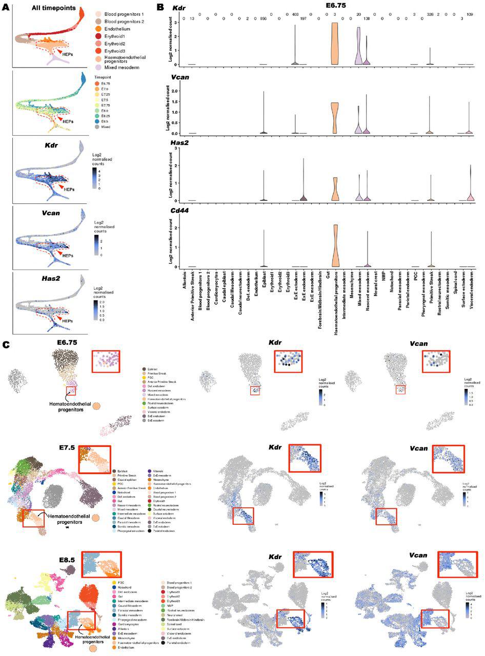
    (A) Violin plots for Hapln1-4 expression by cells of various lineages from E6.75 embryos. No expression of Hapln1-4 is detected in hematoendothelial progenitors (red arrowhead) at this stage. (B) HAPLN1 staining was not detected in E7.5 blood islands marked by versican staining (white arrowheads). (C) ADAMTS-cleaved versican staining (anti-DPEAAE) is undetectable in E7.5 blood islands (white arrowheads). Scale bars = 100μm in B and C .

    Journal: bioRxiv

    Article Title: The versican-hyaluronan complex provides an essential extracellular matrix niche for Flk1+ hematoendothelial progenitors

    doi: 10.1101/753418

    Figure Lengend Snippet: (A) Violin plots for Hapln1-4 expression by cells of various lineages from E6.75 embryos. No expression of Hapln1-4 is detected in hematoendothelial progenitors (red arrowhead) at this stage. (B) HAPLN1 staining was not detected in E7.5 blood islands marked by versican staining (white arrowheads). (C) ADAMTS-cleaved versican staining (anti-DPEAAE) is undetectable in E7.5 blood islands (white arrowheads). Scale bars = 100μm in B and C .

    Article Snippet: For staining whole mount embryos, yolk sacs, vibratome sections and cell culture chamber slides the following antibodies, staining reagents and dilutions were used: Rabbit polyclonal anti-mouse versican GAG β domain (Millipore-Sigma, catalog no. AB1033) 1:200; Rabbit polyclonal anti-versican cleavage site (anti-Vc) [ ] 1:400; Rabbit polyclonal anti-versican V 0 /V 1 neo cleavage antibody (DPEAAE) (Thermo Fisher, catalog no. PA1-1748A) 1:400; biotinylated hyaluronan binding protein (HAbp) (Millipore-Sigma, Calbiochem, catalog no. 385911) 1:100; rabbit polyclonal anti-fibronectin (Abcam, catalog no. Ab2413) 1:200; rabbit polyclonal anti-collagen-IV (Rockland antibodies and assays, catalog no. 600-401-106) 1:400; rat monoclonal anti-mouse Flk1 (clone Avas12, Thermo Fisher, catalog no. 17-5821-81) 1:200; hamster anti-mouse CD31 (Milipore-Sigma, catalog no. MAB1398Z) 1:400; FITC-conjugated anti-mouse CD41 (Biolegend, catalog no.133904) 1:100; mouse monoclonal Cy3 conjugated smooth muscle α -actin (Sigma-Aldrich, catalog no. C6198) 1:600;Alexa Fluor-568 phalloidin (Life Technologies, catalog no. A12380) 1:500; Streptavidin-FITC (Invitrogen, catalog no. SA1001) 1:400; Goat anti-mouse Ihh N-terminus (R&D Systems, catalog no. AF1705) 1:400; Mouse anti-Hapln1 (Link protein) (DSHB, 9/30/8-A-4-C) 1:100.

    Techniques: Expressing, Staining

    (A) Versican (green) and HAbp (red) staining of E8.5 wild type and Vcan hdf/hdf embryos showing severely reduced HAbp staining in Vcan hdf/hdf embryos. Arrowheads indicate versican and HAbp-stained yolk sac blood islands. (B) (Top) Fluorophore-assisted carbohydrate electrophoresis (FACE) analysis for chondroitin sulfate (CS-FACE) and hyaluronan (HA-FACE) in E8.5 littermate embryos. Versican is the major CS-proteoglycan in E8.5 embryos. HA is significantly reduced in Vcan hdf/hdf embryos. (Bottom) Quantification of CS and HA FACE, normalized to DNA (n=4 embryos from each genotype, error bars= S.E.M.,**, p<0.01; ***, p<0.001; ****, p<0.0001). (C) 1 μm thick, toluidine blue-stained Eponate 12 sections from E8.5 wild type and Vcan hdf/hdf embryos show compaction of craniofacial mesenchymal cells (Me) and loss of cardiac jelly (CJ) between the myocardium (Mc) and endocardium (En). The red line indicates the boundary of neural epithelium (NE) with mesenchyme. (D) E8.5 wild Vcan hdf/hdf embryo sections stained with F-actin (red) and fibronectin (green) show stronger fibronectin staining. (E) qRT-PCR shows increased Tmem2 transcription in Vcan hdf/hdf embryos and yolk sacs (N=3 embryos and yolk sacs from each genotype, error bars= S.E.M.;***, p<0.001; ****, p<0.0001). Scale bar in A = 200μm, 50μm and 20μm in C .

    Journal: bioRxiv

    Article Title: The versican-hyaluronan complex provides an essential extracellular matrix niche for Flk1+ hematoendothelial progenitors

    doi: 10.1101/753418

    Figure Lengend Snippet: (A) Versican (green) and HAbp (red) staining of E8.5 wild type and Vcan hdf/hdf embryos showing severely reduced HAbp staining in Vcan hdf/hdf embryos. Arrowheads indicate versican and HAbp-stained yolk sac blood islands. (B) (Top) Fluorophore-assisted carbohydrate electrophoresis (FACE) analysis for chondroitin sulfate (CS-FACE) and hyaluronan (HA-FACE) in E8.5 littermate embryos. Versican is the major CS-proteoglycan in E8.5 embryos. HA is significantly reduced in Vcan hdf/hdf embryos. (Bottom) Quantification of CS and HA FACE, normalized to DNA (n=4 embryos from each genotype, error bars= S.E.M.,**, p<0.01; ***, p<0.001; ****, p<0.0001). (C) 1 μm thick, toluidine blue-stained Eponate 12 sections from E8.5 wild type and Vcan hdf/hdf embryos show compaction of craniofacial mesenchymal cells (Me) and loss of cardiac jelly (CJ) between the myocardium (Mc) and endocardium (En). The red line indicates the boundary of neural epithelium (NE) with mesenchyme. (D) E8.5 wild Vcan hdf/hdf embryo sections stained with F-actin (red) and fibronectin (green) show stronger fibronectin staining. (E) qRT-PCR shows increased Tmem2 transcription in Vcan hdf/hdf embryos and yolk sacs (N=3 embryos and yolk sacs from each genotype, error bars= S.E.M.;***, p<0.001; ****, p<0.0001). Scale bar in A = 200μm, 50μm and 20μm in C .

    Article Snippet: For staining whole mount embryos, yolk sacs, vibratome sections and cell culture chamber slides the following antibodies, staining reagents and dilutions were used: Rabbit polyclonal anti-mouse versican GAG β domain (Millipore-Sigma, catalog no. AB1033) 1:200; Rabbit polyclonal anti-versican cleavage site (anti-Vc) [ ] 1:400; Rabbit polyclonal anti-versican V 0 /V 1 neo cleavage antibody (DPEAAE) (Thermo Fisher, catalog no. PA1-1748A) 1:400; biotinylated hyaluronan binding protein (HAbp) (Millipore-Sigma, Calbiochem, catalog no. 385911) 1:100; rabbit polyclonal anti-fibronectin (Abcam, catalog no. Ab2413) 1:200; rabbit polyclonal anti-collagen-IV (Rockland antibodies and assays, catalog no. 600-401-106) 1:400; rat monoclonal anti-mouse Flk1 (clone Avas12, Thermo Fisher, catalog no. 17-5821-81) 1:200; hamster anti-mouse CD31 (Milipore-Sigma, catalog no. MAB1398Z) 1:400; FITC-conjugated anti-mouse CD41 (Biolegend, catalog no.133904) 1:100; mouse monoclonal Cy3 conjugated smooth muscle α -actin (Sigma-Aldrich, catalog no. C6198) 1:600;Alexa Fluor-568 phalloidin (Life Technologies, catalog no. A12380) 1:500; Streptavidin-FITC (Invitrogen, catalog no. SA1001) 1:400; Goat anti-mouse Ihh N-terminus (R&D Systems, catalog no. AF1705) 1:400; Mouse anti-Hapln1 (Link protein) (DSHB, 9/30/8-A-4-C) 1:100.

    Techniques: Staining, Electrophoresis, Quantitative RT-PCR

    (A) E9.5 Has1+3 null embryos (control) and Has1+2+3 null embryos showing the dilated pericardial sac in the latter (arrowhead) similar to Vcan hdf/hdf . (B) Has1+3 null yolk sac (control) and Has1+2+3 null yolk sac co-stained with HAbp (green) and versican or cleaved versican (DPEAAE) (red) show loss of both HA and versican although cleaved versican (DPEAAE, red, bottom) is present in Has1+2+3 null yolk sac. (C-D) Has1+3 null yolk sac (control) and Has1+2+3 null embryos co-stained with HAbp and versican (C) or cleaved versican (DPEAAE) (D) , show loss of HA and versican staining and weaker DPEAAE staining in the Has1+2+3 null embryos. Scale bars = 200μm in in C , D .

    Journal: bioRxiv

    Article Title: The versican-hyaluronan complex provides an essential extracellular matrix niche for Flk1+ hematoendothelial progenitors

    doi: 10.1101/753418

    Figure Lengend Snippet: (A) E9.5 Has1+3 null embryos (control) and Has1+2+3 null embryos showing the dilated pericardial sac in the latter (arrowhead) similar to Vcan hdf/hdf . (B) Has1+3 null yolk sac (control) and Has1+2+3 null yolk sac co-stained with HAbp (green) and versican or cleaved versican (DPEAAE) (red) show loss of both HA and versican although cleaved versican (DPEAAE, red, bottom) is present in Has1+2+3 null yolk sac. (C-D) Has1+3 null yolk sac (control) and Has1+2+3 null embryos co-stained with HAbp and versican (C) or cleaved versican (DPEAAE) (D) , show loss of HA and versican staining and weaker DPEAAE staining in the Has1+2+3 null embryos. Scale bars = 200μm in in C , D .

    Article Snippet: For staining whole mount embryos, yolk sacs, vibratome sections and cell culture chamber slides the following antibodies, staining reagents and dilutions were used: Rabbit polyclonal anti-mouse versican GAG β domain (Millipore-Sigma, catalog no. AB1033) 1:200; Rabbit polyclonal anti-versican cleavage site (anti-Vc) [ ] 1:400; Rabbit polyclonal anti-versican V 0 /V 1 neo cleavage antibody (DPEAAE) (Thermo Fisher, catalog no. PA1-1748A) 1:400; biotinylated hyaluronan binding protein (HAbp) (Millipore-Sigma, Calbiochem, catalog no. 385911) 1:100; rabbit polyclonal anti-fibronectin (Abcam, catalog no. Ab2413) 1:200; rabbit polyclonal anti-collagen-IV (Rockland antibodies and assays, catalog no. 600-401-106) 1:400; rat monoclonal anti-mouse Flk1 (clone Avas12, Thermo Fisher, catalog no. 17-5821-81) 1:200; hamster anti-mouse CD31 (Milipore-Sigma, catalog no. MAB1398Z) 1:400; FITC-conjugated anti-mouse CD41 (Biolegend, catalog no.133904) 1:100; mouse monoclonal Cy3 conjugated smooth muscle α -actin (Sigma-Aldrich, catalog no. C6198) 1:600;Alexa Fluor-568 phalloidin (Life Technologies, catalog no. A12380) 1:500; Streptavidin-FITC (Invitrogen, catalog no. SA1001) 1:400; Goat anti-mouse Ihh N-terminus (R&D Systems, catalog no. AF1705) 1:400; Mouse anti-Hapln1 (Link protein) (DSHB, 9/30/8-A-4-C) 1:100.

    Techniques: Staining

    (A) Whole mount images of Has2 Fl/Fl ; Sox2 Cre yolk sacs and embryos showing loss of yolk sac vasculature and heart defect (red arrowhead) compared to control littermates (N=8 each genotype). (B) RNAScope in situ hybridization for Has2, Vcan exon 7 (GAG α ) and exon 8 (GAG β ) shows residual Has2 transcript and slightly reduced Vcan transcript labeling in Has2 Fl/Fl ; Sox2 Cre embryos (n=4 embryos each probe). (C) Has2 Fl/Fl ; Sox2 Cre yolk sacs show loss of HA and versican staining (upper panels) and increased versican catabolism is revealed by DPEAAE staining (lower panels) (N=4 each group). (D) Has2 Fl/Fl ; Sox2 Cre embryos show loss of HA and versican staining (N=3 each group). (E) Has2 Fl/Fl ; Sox2 Cre embryos show increased DPEAAE staining (N=3 each group).

    Journal: bioRxiv

    Article Title: The versican-hyaluronan complex provides an essential extracellular matrix niche for Flk1+ hematoendothelial progenitors

    doi: 10.1101/753418

    Figure Lengend Snippet: (A) Whole mount images of Has2 Fl/Fl ; Sox2 Cre yolk sacs and embryos showing loss of yolk sac vasculature and heart defect (red arrowhead) compared to control littermates (N=8 each genotype). (B) RNAScope in situ hybridization for Has2, Vcan exon 7 (GAG α ) and exon 8 (GAG β ) shows residual Has2 transcript and slightly reduced Vcan transcript labeling in Has2 Fl/Fl ; Sox2 Cre embryos (n=4 embryos each probe). (C) Has2 Fl/Fl ; Sox2 Cre yolk sacs show loss of HA and versican staining (upper panels) and increased versican catabolism is revealed by DPEAAE staining (lower panels) (N=4 each group). (D) Has2 Fl/Fl ; Sox2 Cre embryos show loss of HA and versican staining (N=3 each group). (E) Has2 Fl/Fl ; Sox2 Cre embryos show increased DPEAAE staining (N=3 each group).

    Article Snippet: For staining whole mount embryos, yolk sacs, vibratome sections and cell culture chamber slides the following antibodies, staining reagents and dilutions were used: Rabbit polyclonal anti-mouse versican GAG β domain (Millipore-Sigma, catalog no. AB1033) 1:200; Rabbit polyclonal anti-versican cleavage site (anti-Vc) [ ] 1:400; Rabbit polyclonal anti-versican V 0 /V 1 neo cleavage antibody (DPEAAE) (Thermo Fisher, catalog no. PA1-1748A) 1:400; biotinylated hyaluronan binding protein (HAbp) (Millipore-Sigma, Calbiochem, catalog no. 385911) 1:100; rabbit polyclonal anti-fibronectin (Abcam, catalog no. Ab2413) 1:200; rabbit polyclonal anti-collagen-IV (Rockland antibodies and assays, catalog no. 600-401-106) 1:400; rat monoclonal anti-mouse Flk1 (clone Avas12, Thermo Fisher, catalog no. 17-5821-81) 1:200; hamster anti-mouse CD31 (Milipore-Sigma, catalog no. MAB1398Z) 1:400; FITC-conjugated anti-mouse CD41 (Biolegend, catalog no.133904) 1:100; mouse monoclonal Cy3 conjugated smooth muscle α -actin (Sigma-Aldrich, catalog no. C6198) 1:600;Alexa Fluor-568 phalloidin (Life Technologies, catalog no. A12380) 1:500; Streptavidin-FITC (Invitrogen, catalog no. SA1001) 1:400; Goat anti-mouse Ihh N-terminus (R&D Systems, catalog no. AF1705) 1:400; Mouse anti-Hapln1 (Link protein) (DSHB, 9/30/8-A-4-C) 1:100.

    Techniques: In Situ Hybridization, Labeling, Staining

    (A) Vcan locus showing targeting by independent guide RNAs (gRNAs) targeting exons 2 and 3. Scissors indicate the Cas9 cleavage site 3 bp from the protospacer adjacent motif (PAM, orange lettering). An exon 2 mutant mESC clone (D8) had a homozygous 1 bp insertion (bold red letters) and an exon 3 mutant clone (F9) had heterozygous targeting: deletion of T/A in one allele and a 19-bp deletion in the other resulting in frame-shifts. (B) Western blot of 10-day differentiated EBs shows no versican in EBs derived from clones D8 and F9. (C) qRT-PCR analysis showing unaffected expression of pluripotency markers Oct4 , Sox2 , Nanog and C-myc in D8 and F9 (n=3 independent batches of EBs from each genotype, error bars= S.E.). (D) 4-day-old Vcan -null embryoid bodies embedded in collagen I and treated with VEGF 165 for 12 days lack vascular sprouting identified by CD31 (green) or α -SMA (red) immunostaining (n=3 independent batches of EBs from each genotype). (E) D8 and F9 EBs show significantly fewer vascular sprouts (n=3 independent batches of EBs from each genotype, error bars= S.E. **, p<0.01; ***, p<0.001). (F) Fewer sprouts/EB were seen in D8 and F9 EBs. (n=3 independent batches of EBs from each genotype, error bars= S.D. ***, p<0.001; ****, p<0.0001). (G) Significantly shorter sprouts in Vcan null lines (n=3 independent batches of EBs from each genotype, error bars= S.D. ****, p<0.0001). (H) Methylcellulose colony assay using dissociated 10-day old EBs shows significantly fewer colonies in Vcan null EBs. (N=3 independent batches of EBs from each genotype, error bars=S.D. **, p<0.01). Scale bar in D is 200 μm.

    Journal: bioRxiv

    Article Title: The versican-hyaluronan complex provides an essential extracellular matrix niche for Flk1+ hematoendothelial progenitors

    doi: 10.1101/753418

    Figure Lengend Snippet: (A) Vcan locus showing targeting by independent guide RNAs (gRNAs) targeting exons 2 and 3. Scissors indicate the Cas9 cleavage site 3 bp from the protospacer adjacent motif (PAM, orange lettering). An exon 2 mutant mESC clone (D8) had a homozygous 1 bp insertion (bold red letters) and an exon 3 mutant clone (F9) had heterozygous targeting: deletion of T/A in one allele and a 19-bp deletion in the other resulting in frame-shifts. (B) Western blot of 10-day differentiated EBs shows no versican in EBs derived from clones D8 and F9. (C) qRT-PCR analysis showing unaffected expression of pluripotency markers Oct4 , Sox2 , Nanog and C-myc in D8 and F9 (n=3 independent batches of EBs from each genotype, error bars= S.E.). (D) 4-day-old Vcan -null embryoid bodies embedded in collagen I and treated with VEGF 165 for 12 days lack vascular sprouting identified by CD31 (green) or α -SMA (red) immunostaining (n=3 independent batches of EBs from each genotype). (E) D8 and F9 EBs show significantly fewer vascular sprouts (n=3 independent batches of EBs from each genotype, error bars= S.E. **, p<0.01; ***, p<0.001). (F) Fewer sprouts/EB were seen in D8 and F9 EBs. (n=3 independent batches of EBs from each genotype, error bars= S.D. ***, p<0.001; ****, p<0.0001). (G) Significantly shorter sprouts in Vcan null lines (n=3 independent batches of EBs from each genotype, error bars= S.D. ****, p<0.0001). (H) Methylcellulose colony assay using dissociated 10-day old EBs shows significantly fewer colonies in Vcan null EBs. (N=3 independent batches of EBs from each genotype, error bars=S.D. **, p<0.01). Scale bar in D is 200 μm.

    Article Snippet: For staining whole mount embryos, yolk sacs, vibratome sections and cell culture chamber slides the following antibodies, staining reagents and dilutions were used: Rabbit polyclonal anti-mouse versican GAG β domain (Millipore-Sigma, catalog no. AB1033) 1:200; Rabbit polyclonal anti-versican cleavage site (anti-Vc) [ ] 1:400; Rabbit polyclonal anti-versican V 0 /V 1 neo cleavage antibody (DPEAAE) (Thermo Fisher, catalog no. PA1-1748A) 1:400; biotinylated hyaluronan binding protein (HAbp) (Millipore-Sigma, Calbiochem, catalog no. 385911) 1:100; rabbit polyclonal anti-fibronectin (Abcam, catalog no. Ab2413) 1:200; rabbit polyclonal anti-collagen-IV (Rockland antibodies and assays, catalog no. 600-401-106) 1:400; rat monoclonal anti-mouse Flk1 (clone Avas12, Thermo Fisher, catalog no. 17-5821-81) 1:200; hamster anti-mouse CD31 (Milipore-Sigma, catalog no. MAB1398Z) 1:400; FITC-conjugated anti-mouse CD41 (Biolegend, catalog no.133904) 1:100; mouse monoclonal Cy3 conjugated smooth muscle α -actin (Sigma-Aldrich, catalog no. C6198) 1:600;Alexa Fluor-568 phalloidin (Life Technologies, catalog no. A12380) 1:500; Streptavidin-FITC (Invitrogen, catalog no. SA1001) 1:400; Goat anti-mouse Ihh N-terminus (R&D Systems, catalog no. AF1705) 1:400; Mouse anti-Hapln1 (Link protein) (DSHB, 9/30/8-A-4-C) 1:100.

    Techniques: Mutagenesis, Western Blot, Derivative Assay, Clone Assay, Quantitative RT-PCR, Expressing, Immunostaining, Colony Assay

    (A-F) 10-day differentiated embryoid bodies from three independent experiments were analyzed by qRT-PCR. Has2 transcription in embryoid bodies was unaffected by the loss of versican. mRNA for the mesoderm marker Brachyury was significantly lower in F9 but not in D8 embryoid bodies contrasting with Vcan hdf/hdf embryos (main ). Expression of the hematovascular progenitor marker Flk1, blood lineage commitment progenitor markers Itga2b (CD41) and Runx1 , differentiated blood markers Hbb ( β -globin), Gata1 and Ptprc (CD45) and endothelial marker Pecam1 (CD31) was reduced in Vcan -null embryoid bodies (N=3 independent batches of 10 day embryoid bodies from each genotype, error bars= S.E.M.,*, p<0.05; **, p<0.01; ***, p<0.0001).

    Journal: bioRxiv

    Article Title: The versican-hyaluronan complex provides an essential extracellular matrix niche for Flk1+ hematoendothelial progenitors

    doi: 10.1101/753418

    Figure Lengend Snippet: (A-F) 10-day differentiated embryoid bodies from three independent experiments were analyzed by qRT-PCR. Has2 transcription in embryoid bodies was unaffected by the loss of versican. mRNA for the mesoderm marker Brachyury was significantly lower in F9 but not in D8 embryoid bodies contrasting with Vcan hdf/hdf embryos (main ). Expression of the hematovascular progenitor marker Flk1, blood lineage commitment progenitor markers Itga2b (CD41) and Runx1 , differentiated blood markers Hbb ( β -globin), Gata1 and Ptprc (CD45) and endothelial marker Pecam1 (CD31) was reduced in Vcan -null embryoid bodies (N=3 independent batches of 10 day embryoid bodies from each genotype, error bars= S.E.M.,*, p<0.05; **, p<0.01; ***, p<0.0001).

    Article Snippet: For staining whole mount embryos, yolk sacs, vibratome sections and cell culture chamber slides the following antibodies, staining reagents and dilutions were used: Rabbit polyclonal anti-mouse versican GAG β domain (Millipore-Sigma, catalog no. AB1033) 1:200; Rabbit polyclonal anti-versican cleavage site (anti-Vc) [ ] 1:400; Rabbit polyclonal anti-versican V 0 /V 1 neo cleavage antibody (DPEAAE) (Thermo Fisher, catalog no. PA1-1748A) 1:400; biotinylated hyaluronan binding protein (HAbp) (Millipore-Sigma, Calbiochem, catalog no. 385911) 1:100; rabbit polyclonal anti-fibronectin (Abcam, catalog no. Ab2413) 1:200; rabbit polyclonal anti-collagen-IV (Rockland antibodies and assays, catalog no. 600-401-106) 1:400; rat monoclonal anti-mouse Flk1 (clone Avas12, Thermo Fisher, catalog no. 17-5821-81) 1:200; hamster anti-mouse CD31 (Milipore-Sigma, catalog no. MAB1398Z) 1:400; FITC-conjugated anti-mouse CD41 (Biolegend, catalog no.133904) 1:100; mouse monoclonal Cy3 conjugated smooth muscle α -actin (Sigma-Aldrich, catalog no. C6198) 1:600;Alexa Fluor-568 phalloidin (Life Technologies, catalog no. A12380) 1:500; Streptavidin-FITC (Invitrogen, catalog no. SA1001) 1:400; Goat anti-mouse Ihh N-terminus (R&D Systems, catalog no. AF1705) 1:400; Mouse anti-Hapln1 (Link protein) (DSHB, 9/30/8-A-4-C) 1:100.

    Techniques: Quantitative RT-PCR, Marker, Expressing

    (A) Increased versican (red) and HAbp staining (green) in ADAMTS9 -deficient (D12) RPE-1 cells. Versican and HA co-stained cables are formed in D12 cells. (B) qRT-PCR showing no change in HAS1-3 or CD44 but dramatically reduced TMEM2 expression in D12 cells. (n=3 independent RNA extractions, error bars= S.E.M., *, p<0.05; ****, p<0.0001). (C) VCAN knockdown reduces both versican (red) and HA staining in D12 cells. (D) Recombinant VEGF 165 or Ihh (green) co-staining with versican (red) in control siRNA-transfected D12 cells is lost in VCAN siRNA-transfected D12 cells. (E) The schematic illustrates chondroitinase ABC removal of versican GAG chains and the experimental timeline used. (F) Fluorescence microscope of VEGF and Ihh with versican, with or without chondroitinase ABC treatment prior to addition of recombinant VEGF 165 or Ihh (green) shows reduced binding of both growth factors after CS-chain removal, but does not affect versican core protein staining (red).

    Journal: bioRxiv

    Article Title: The versican-hyaluronan complex provides an essential extracellular matrix niche for Flk1+ hematoendothelial progenitors

    doi: 10.1101/753418

    Figure Lengend Snippet: (A) Increased versican (red) and HAbp staining (green) in ADAMTS9 -deficient (D12) RPE-1 cells. Versican and HA co-stained cables are formed in D12 cells. (B) qRT-PCR showing no change in HAS1-3 or CD44 but dramatically reduced TMEM2 expression in D12 cells. (n=3 independent RNA extractions, error bars= S.E.M., *, p<0.05; ****, p<0.0001). (C) VCAN knockdown reduces both versican (red) and HA staining in D12 cells. (D) Recombinant VEGF 165 or Ihh (green) co-staining with versican (red) in control siRNA-transfected D12 cells is lost in VCAN siRNA-transfected D12 cells. (E) The schematic illustrates chondroitinase ABC removal of versican GAG chains and the experimental timeline used. (F) Fluorescence microscope of VEGF and Ihh with versican, with or without chondroitinase ABC treatment prior to addition of recombinant VEGF 165 or Ihh (green) shows reduced binding of both growth factors after CS-chain removal, but does not affect versican core protein staining (red).

    Article Snippet: For staining whole mount embryos, yolk sacs, vibratome sections and cell culture chamber slides the following antibodies, staining reagents and dilutions were used: Rabbit polyclonal anti-mouse versican GAG β domain (Millipore-Sigma, catalog no. AB1033) 1:200; Rabbit polyclonal anti-versican cleavage site (anti-Vc) [ ] 1:400; Rabbit polyclonal anti-versican V 0 /V 1 neo cleavage antibody (DPEAAE) (Thermo Fisher, catalog no. PA1-1748A) 1:400; biotinylated hyaluronan binding protein (HAbp) (Millipore-Sigma, Calbiochem, catalog no. 385911) 1:100; rabbit polyclonal anti-fibronectin (Abcam, catalog no. Ab2413) 1:200; rabbit polyclonal anti-collagen-IV (Rockland antibodies and assays, catalog no. 600-401-106) 1:400; rat monoclonal anti-mouse Flk1 (clone Avas12, Thermo Fisher, catalog no. 17-5821-81) 1:200; hamster anti-mouse CD31 (Milipore-Sigma, catalog no. MAB1398Z) 1:400; FITC-conjugated anti-mouse CD41 (Biolegend, catalog no.133904) 1:100; mouse monoclonal Cy3 conjugated smooth muscle α -actin (Sigma-Aldrich, catalog no. C6198) 1:600;Alexa Fluor-568 phalloidin (Life Technologies, catalog no. A12380) 1:500; Streptavidin-FITC (Invitrogen, catalog no. SA1001) 1:400; Goat anti-mouse Ihh N-terminus (R&D Systems, catalog no. AF1705) 1:400; Mouse anti-Hapln1 (Link protein) (DSHB, 9/30/8-A-4-C) 1:100.

    Techniques: Staining, Quantitative RT-PCR, Expressing, Recombinant, Transfection, Fluorescence, Microscopy, Binding Assay

    (A) Four independent representative areas of wild type RPE-1 and ADAMTS9 deficient RPE-1 (D12) cell cultures stained with HAbp showing HA accumulation and numerous long HA-cables present in D12 cultures, but not parental RPE1 cells (white arrowheads). (B) High-magnification confocal images showing two examples of long HA cables (green) decorated with versican (red). Scale bars = 50μm in A and 5μm in B .

    Journal: bioRxiv

    Article Title: The versican-hyaluronan complex provides an essential extracellular matrix niche for Flk1+ hematoendothelial progenitors

    doi: 10.1101/753418

    Figure Lengend Snippet: (A) Four independent representative areas of wild type RPE-1 and ADAMTS9 deficient RPE-1 (D12) cell cultures stained with HAbp showing HA accumulation and numerous long HA-cables present in D12 cultures, but not parental RPE1 cells (white arrowheads). (B) High-magnification confocal images showing two examples of long HA cables (green) decorated with versican (red). Scale bars = 50μm in A and 5μm in B .

    Article Snippet: For staining whole mount embryos, yolk sacs, vibratome sections and cell culture chamber slides the following antibodies, staining reagents and dilutions were used: Rabbit polyclonal anti-mouse versican GAG β domain (Millipore-Sigma, catalog no. AB1033) 1:200; Rabbit polyclonal anti-versican cleavage site (anti-Vc) [ ] 1:400; Rabbit polyclonal anti-versican V 0 /V 1 neo cleavage antibody (DPEAAE) (Thermo Fisher, catalog no. PA1-1748A) 1:400; biotinylated hyaluronan binding protein (HAbp) (Millipore-Sigma, Calbiochem, catalog no. 385911) 1:100; rabbit polyclonal anti-fibronectin (Abcam, catalog no. Ab2413) 1:200; rabbit polyclonal anti-collagen-IV (Rockland antibodies and assays, catalog no. 600-401-106) 1:400; rat monoclonal anti-mouse Flk1 (clone Avas12, Thermo Fisher, catalog no. 17-5821-81) 1:200; hamster anti-mouse CD31 (Milipore-Sigma, catalog no. MAB1398Z) 1:400; FITC-conjugated anti-mouse CD41 (Biolegend, catalog no.133904) 1:100; mouse monoclonal Cy3 conjugated smooth muscle α -actin (Sigma-Aldrich, catalog no. C6198) 1:600;Alexa Fluor-568 phalloidin (Life Technologies, catalog no. A12380) 1:500; Streptavidin-FITC (Invitrogen, catalog no. SA1001) 1:400; Goat anti-mouse Ihh N-terminus (R&D Systems, catalog no. AF1705) 1:400; Mouse anti-Hapln1 (Link protein) (DSHB, 9/30/8-A-4-C) 1:100.

    Techniques: Staining

    (A) Wild type RPE-1 and ADAMTS9 -deficient RPE-1 (D12) cells treated with PBS or increasing concentrations of recombinant biotinylated VEGF 165 (green) showing dose-dependent VEGF staining intensity and colocalization with versican (red) in D12 cells. (B) Super-resolution confocal images of a long versican/HA cable showing patches of VEGF 165 (green) deposited along the versican stained cable (red). Nuclei are stained blue with DAPI. The upper merged panel shows some strongly co-staining regions (arrowheads), whereas the center panel shows co-staining in the X-Z plane. The lower panels show the single-color images of the areas marked as Box 1 and Box 2 in the upper panel. Scale bars = 50μm in A and 10μm in B .

    Journal: bioRxiv

    Article Title: The versican-hyaluronan complex provides an essential extracellular matrix niche for Flk1+ hematoendothelial progenitors

    doi: 10.1101/753418

    Figure Lengend Snippet: (A) Wild type RPE-1 and ADAMTS9 -deficient RPE-1 (D12) cells treated with PBS or increasing concentrations of recombinant biotinylated VEGF 165 (green) showing dose-dependent VEGF staining intensity and colocalization with versican (red) in D12 cells. (B) Super-resolution confocal images of a long versican/HA cable showing patches of VEGF 165 (green) deposited along the versican stained cable (red). Nuclei are stained blue with DAPI. The upper merged panel shows some strongly co-staining regions (arrowheads), whereas the center panel shows co-staining in the X-Z plane. The lower panels show the single-color images of the areas marked as Box 1 and Box 2 in the upper panel. Scale bars = 50μm in A and 10μm in B .

    Article Snippet: For staining whole mount embryos, yolk sacs, vibratome sections and cell culture chamber slides the following antibodies, staining reagents and dilutions were used: Rabbit polyclonal anti-mouse versican GAG β domain (Millipore-Sigma, catalog no. AB1033) 1:200; Rabbit polyclonal anti-versican cleavage site (anti-Vc) [ ] 1:400; Rabbit polyclonal anti-versican V 0 /V 1 neo cleavage antibody (DPEAAE) (Thermo Fisher, catalog no. PA1-1748A) 1:400; biotinylated hyaluronan binding protein (HAbp) (Millipore-Sigma, Calbiochem, catalog no. 385911) 1:100; rabbit polyclonal anti-fibronectin (Abcam, catalog no. Ab2413) 1:200; rabbit polyclonal anti-collagen-IV (Rockland antibodies and assays, catalog no. 600-401-106) 1:400; rat monoclonal anti-mouse Flk1 (clone Avas12, Thermo Fisher, catalog no. 17-5821-81) 1:200; hamster anti-mouse CD31 (Milipore-Sigma, catalog no. MAB1398Z) 1:400; FITC-conjugated anti-mouse CD41 (Biolegend, catalog no.133904) 1:100; mouse monoclonal Cy3 conjugated smooth muscle α -actin (Sigma-Aldrich, catalog no. C6198) 1:600;Alexa Fluor-568 phalloidin (Life Technologies, catalog no. A12380) 1:500; Streptavidin-FITC (Invitrogen, catalog no. SA1001) 1:400; Goat anti-mouse Ihh N-terminus (R&D Systems, catalog no. AF1705) 1:400; Mouse anti-Hapln1 (Link protein) (DSHB, 9/30/8-A-4-C) 1:100.

    Techniques: Recombinant, Staining

    (A) Wild type RPE-1 and ADAMTS9 deficient (D12) cells treated with PBS or increasing concentrations of recombinant Ihh N-terminal domain (green) showing dose-dependent deposition and increased co-staining (yellow) with versican (red) in D12 cells. Nuclei are stained blue with DAPI. (B) Super-resolution confocal image of a versican-HA stained cable showing Ihh staining (green) along a versican stained cable (red). Strongly co-stained regions are indicated by arrowheads. The center panel shows the Y-Z plane. Bottom panels show single-color images of the area imaged in the upper panel. Scale bars = 20μm in A and 10μm in B .

    Journal: bioRxiv

    Article Title: The versican-hyaluronan complex provides an essential extracellular matrix niche for Flk1+ hematoendothelial progenitors

    doi: 10.1101/753418

    Figure Lengend Snippet: (A) Wild type RPE-1 and ADAMTS9 deficient (D12) cells treated with PBS or increasing concentrations of recombinant Ihh N-terminal domain (green) showing dose-dependent deposition and increased co-staining (yellow) with versican (red) in D12 cells. Nuclei are stained blue with DAPI. (B) Super-resolution confocal image of a versican-HA stained cable showing Ihh staining (green) along a versican stained cable (red). Strongly co-stained regions are indicated by arrowheads. The center panel shows the Y-Z plane. Bottom panels show single-color images of the area imaged in the upper panel. Scale bars = 20μm in A and 10μm in B .

    Article Snippet: For staining whole mount embryos, yolk sacs, vibratome sections and cell culture chamber slides the following antibodies, staining reagents and dilutions were used: Rabbit polyclonal anti-mouse versican GAG β domain (Millipore-Sigma, catalog no. AB1033) 1:200; Rabbit polyclonal anti-versican cleavage site (anti-Vc) [ ] 1:400; Rabbit polyclonal anti-versican V 0 /V 1 neo cleavage antibody (DPEAAE) (Thermo Fisher, catalog no. PA1-1748A) 1:400; biotinylated hyaluronan binding protein (HAbp) (Millipore-Sigma, Calbiochem, catalog no. 385911) 1:100; rabbit polyclonal anti-fibronectin (Abcam, catalog no. Ab2413) 1:200; rabbit polyclonal anti-collagen-IV (Rockland antibodies and assays, catalog no. 600-401-106) 1:400; rat monoclonal anti-mouse Flk1 (clone Avas12, Thermo Fisher, catalog no. 17-5821-81) 1:200; hamster anti-mouse CD31 (Milipore-Sigma, catalog no. MAB1398Z) 1:400; FITC-conjugated anti-mouse CD41 (Biolegend, catalog no.133904) 1:100; mouse monoclonal Cy3 conjugated smooth muscle α -actin (Sigma-Aldrich, catalog no. C6198) 1:600;Alexa Fluor-568 phalloidin (Life Technologies, catalog no. A12380) 1:500; Streptavidin-FITC (Invitrogen, catalog no. SA1001) 1:400; Goat anti-mouse Ihh N-terminus (R&D Systems, catalog no. AF1705) 1:400; Mouse anti-Hapln1 (Link protein) (DSHB, 9/30/8-A-4-C) 1:100.

    Techniques: Recombinant, Staining

    Journal: eLife

    Article Title: Vascular dimorphism ensured by regulated proteoglycan dynamics favors rapid umbilical artery closure at birth

    doi: 10.7554/eLife.60683

    Figure Lengend Snippet:

    Article Snippet: Antibody , Rabbit polyclonal anti-versican GAG-beta , Millipore Sigma AB1033 , RRID: AB_90462 , IF (1:400) mouse tissue.

    Techniques: RNAscope, In Situ, Software, Gene Expression, Microarray